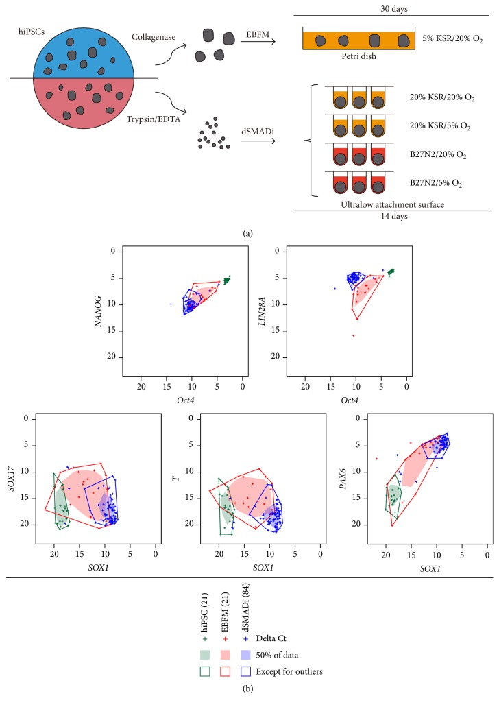
Figure 1.
dSMADi improves the neural induction efficiency of hiPSCs regardless of their somatic tissue origin. (a) Schematic drawing of the neural induction methods used in this study. (b) Bivariate box plots displaying gene expression levels in hiPSCs, day 30 EBFM-derived EBs, and day 14 dSMADi-derived aggregates. Quantitative RT-PCR-generated delta Ct values for the pluripotency marker genes (Oct4, NANOG, and LIN28A), endoderm marker gene (SOX17), mesoderm marker gene (T), and neural marker genes (SOX1 and PAX6) are shown. Green: hiPSCs; red: EBFM; blue: dSMADi. The numbers of clones analyzed are indicated in parentheses and “+” symbols represent each of the delta Ct values. Filled-in regions contain the 50% of the data points, and data points outside of the surrounding lines represent outliers. See also Figures S1 and S2.

