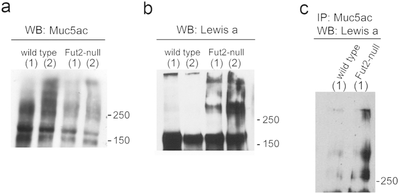
Figure 2. Muc5ac from “non-secretor” Fut2-null mice displays enrichment in Lewis a glycan structures.
Wild-type and Fut2-null mice total gastric mucosa protein lysates were used for evaluation of (a) Muc5ac (45M1) and (b) Lea (SPM279) expression by Western blotting. Labels (1) and (2) represent protein lysates from two independent mice samples. (c) G-sepharose beads coupled with Muc5ac recognizing antibody 45M1 were incubated with wild-type and Fut2-null mice gastric mucosa protein extracts. Proteins that bound to the beads were analyzed by Western blotting with an anti -Lea antibody.

