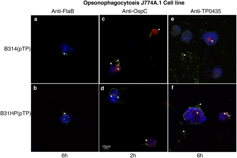
Figure 7. Anti-TP0435 mouse antibodies facilitate only low opsonophagocytosis of B314(pTP) and B31HP(pTP) strains.
(a,b) J774A.1 mouse macrophage cell line failed to phagocytise B31HP(pTP) and B314(pTP) preincubated with antibodies against periplasmic protein FlaB even after 6 h of co-incubation. Extracellular spirochetes (green/yellow marked by arrowhead) were observed on staining of B. burgdorferi with anti-B. burgdorferi OMV antibodies followed by Alexa fluor 488 conjugated anti-mouse antibodies before permeabilization, while to detect intracellular spirochetes, after permeabilization counterstaining was done with anti-OMV antibodies followed by anti-mouse antibodies conjugated to TRITC. (c,d) Preincubation of B314(pTP) and B31HP(pTP) with antibodies against B. burgdorferi surface protein OspC showed phagocytosis by J774A.1 cells within 2 h of co-incubation shown as red intracellular degrading spirochetes. (e,f) B31HP(pTP) and B314(pTP) preincubated with anti-TP0435 antibodies bind to J774A.1 mouse macrophage cell line. After 6 h of co-incubation, both bound or unbound extracellular B. burgdorferi (green marked by arrowhead) and intracellular spirochetes (red marked by arrow) are observed indicating slow but detectable opsonophagocytosis. Scale represents all panels in the figure.

