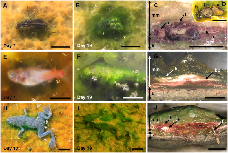
Figure 1. Temporal sequence of the microbial coating of carcasses.
(A–D) Fly (Musca domestica); (E–G) Fish (Paracheirodon innesi), (H–J) Frog (Hymenochirus boettgeri). The left column shows the organisms after one week on the mat. The central column corresponds to carcasses completely covered at day 19. The right column shows the sections cut across microbial mat blocks containing the carcasses. Note the coherence of the sarcophagus around the bodies of flies (C, 5.5 years and D, 8 months), fish (G, 8 months) and frog (J, 12 months). Abbreviations: a, abdomen; b, brain; bb, back bones; h, head; f, femur; mm, microbial mat; s, sediment; t, thorax. (Scale bar: A–D, 5 mm; E–J, 10 mm).

