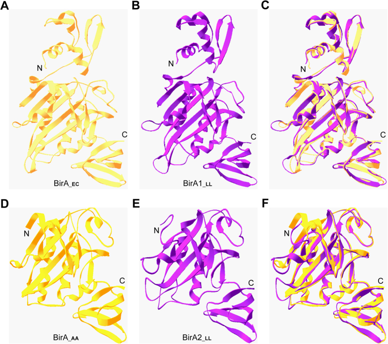
Figure 4. Structural analyses of two BirA homologue proteins from L. lactis.
The E. coli BirA structure (in Panel A) and the modeled structure of L. lactis BirA1 (BirA1_LL) (in Panel B) exhibit strong structural similarity. (C) Structural superposition of BirA1_LL with BirA_EC. The BirA_EC structure (PDB: 1HXD) is in yellow, whereas that of BirA1_LL (with 28.6% identity to BirA_EC structure) is indicated in purple. An overall view of Aquifex aeolicus BirA protein (BirA_AA) structure (in Panel D) and the modeled structure of BirA2_LL protein (in Panel E) (F). Structure superposition of BirA2_LL with BirA_AA. The structure of BirA_AA (PDB: 3EFS) is in gold, whereas the modeled structure of BirA2_LL using BirA_AA as the template with 32.4% identity is highlighted in purple. Designations: AA, Aquifex aeolicus; LL, L. lactis; N, N-terminus; C, C-terminus.

