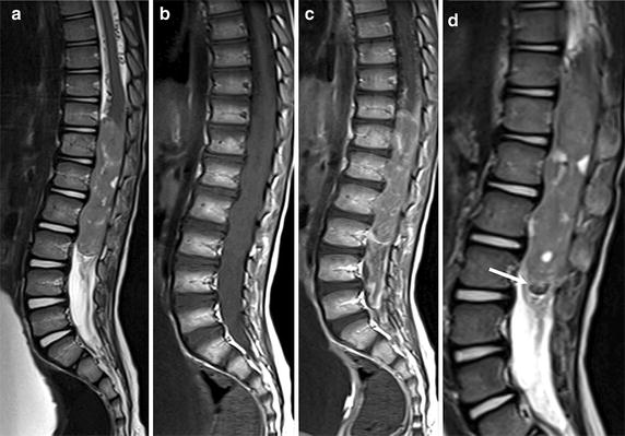
Fig. 1.

Magnetic resonance imaging (MRI) of a 4-year-old boy with spinal glioblastoma multiforme (GBM) of the conus medullaris at the time of diagnosis. MRI shows a large heterogeneous mass extensively filling the spinal canal between T11 and L3. The lesion shows hyperintense and inhomogeneous signal intensity on Sagittal T2-weighted images (a) and isointense signal intensity on Sagittal T1-weighted images (b). After gadolinium (Gd) injection, a diffuse, inhomogeneous enhancement of the tumor is observed, and the tecal sac is filled by abundant enhancing tissue enveloping the conus medullaris and cauda equina (c). The regions of hypointensity within the tumor and along the inferior margin of the lesion shown on sagittal T2-weighted images suggest tumoral bleeding (arrow) (d)
