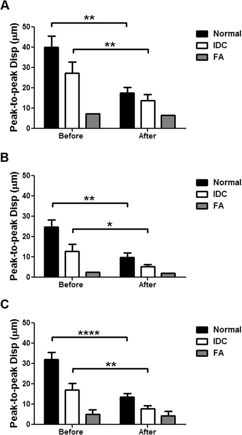
Fig. 3.

Harmonic motion imaging (HMI) displacement change between before and after ablation. a Nine normal, five invasive ductal carcinoma (IDC), and one fibroadenoma (FA) specimens were imaged with the 1D HMI system. b Ten normal, ten IDC, and one FA specimens were imaged with the 2D HMI system. c Combined results with both HMI systems. *p < 0.05, **p < 0.001, and ****p < 0.00001
