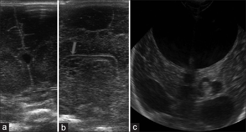
Figure 4.

Variants (a) coronal high resolution ultrasound image demonstrates the cavum septum pellucidum seen as a midline cyst between the frontal horns of lateral ventricles. (b) Two high resolution sagittal images (both sides) shows multiple cysts parallel to the lateral ventricle suggestive of connatal cysts. (c) Coronal image reveals left choroid plexus cyst. Also noted is communicating hydrocephalus
