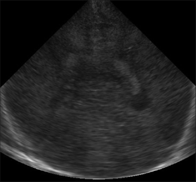
Figure 7.

Hypoxic ischemic encephalopathy. Coronal image demonstrates diffuse hypoxic brain injury as bilaterally symmetric echogenicity with loss of gray-white matter differentiation suggestive of brain edema. CC: Cerebral hemisphere, C: Cerebellum, CP: Choroid plexus; LV: Lateral ventricle
