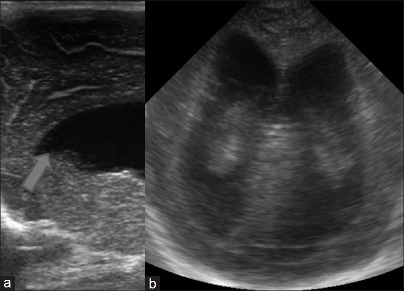
Figure 9.

Intracranial hemorrhage (a) sagittal image shows the typical appearance of germinal matrix hemorrhage as echogenic focus in the caudo-thalamic groove. C: Caudate; TH: Thalamus. (b) Intraventricular hemorrhage is seen on this coronal image as echogenic contents within the frontal horn with layering (arrows). (c) Coronal image demonstrates bilateral temporoparietal echogenic lesions (arrows) suggestive of intraparenchymal hemorrhage. Also note mild mid-line shift towards left side
