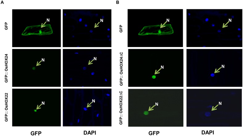
FIGURE 3.
Sub-cellular localization of full-length and truncated (ΔC) OsHOX proteins. (A,B) Sub-cellular localization of full-length (GFP::OsHOX24 and GFP::OsHOX22) (A) and truncated (GFP::OsHOX24ΔC and GFP::OsHOX22ΔC) (B) fusion proteins in onion epidermal cells. Empty GFP vector was used as experimental control. The left panel shows GFP fluorescence followed by DAPI (nucleus (N)-specific dye) staining in the right panel for each construct and vector control.

