FIGURE 4.
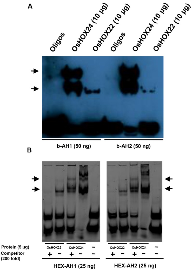
Binding of homeobox TFs to specific DNA (AH1 and AH2) motifs. (A,B) EMSA showing binding of biotinylated AH1 (b-AH1) and AH2 (b-AH2) tetrameric motifs with purified recombinant proteins, 6xHis::OsHOX24 and 6xHis::OsHOX22 (A) and binding of HEX-labeled AH1 and AH2 tetrameric motifs with purified recombinant proteins, 6xHis::OsHOX24 and 6xHis::OsHOX22 (B). For binding reactions, 25–50 nM of annealed oligos were used along with 5–10 μg purified protein and samples were run on 6% native PAGE in 0.25X TBE buffer, followed by development of blot (biotinylated oligos) or direct visualization (HEX-labeled oligos). The arrows indicate the position of binding of recombinant protein with tetrameric motifs as detected by streptavidin-HRP conjugate. For EMSA experiment using HEX-labeled oligos, 200-fold excess of unlabelled oligos were used as competitor in a separate reaction.

