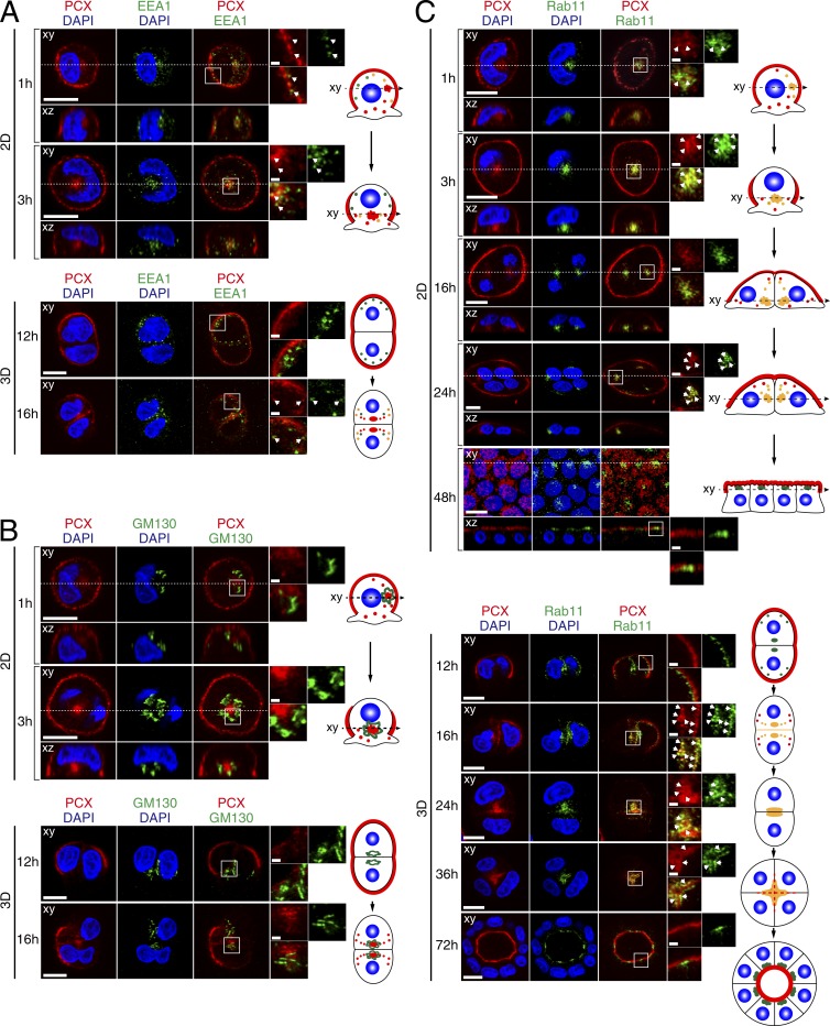
Figure 2.
PCX colocalization with organelle markers in 2D monolayers and in 3D cysts. MDCK II cells were plated on glass bottom dishes (2D) or Matrigel-coated glass slides (3D) and fixed with PFA at the times indicated (see also Fig. S2). (A and B) Cells were costained with anti-PCX antibody (red), DAPI (blue) and an antibody against EEA1 (an early endosome marker; A), GM130 (a Golgi marker; B), or Rab11 (an RE marker; green; C). The confocal xy section (top) and the xz section (bottom), which corresponds to the dashed line in the xy section, of 2D monolayers are shown. The fourth columns show magnifications of the boxed regions in the third columns. The arrows in A show the colocalization points between EEA1 and PCX. Colocalization of PCX with each organelle marker is illustrated schematically on the right side of each panel (red, PCX; green, marker; yellow, colocalization site; and blue, nucleus). Bars: 10 µm; (insets) 1 µm.

