Figure 3.
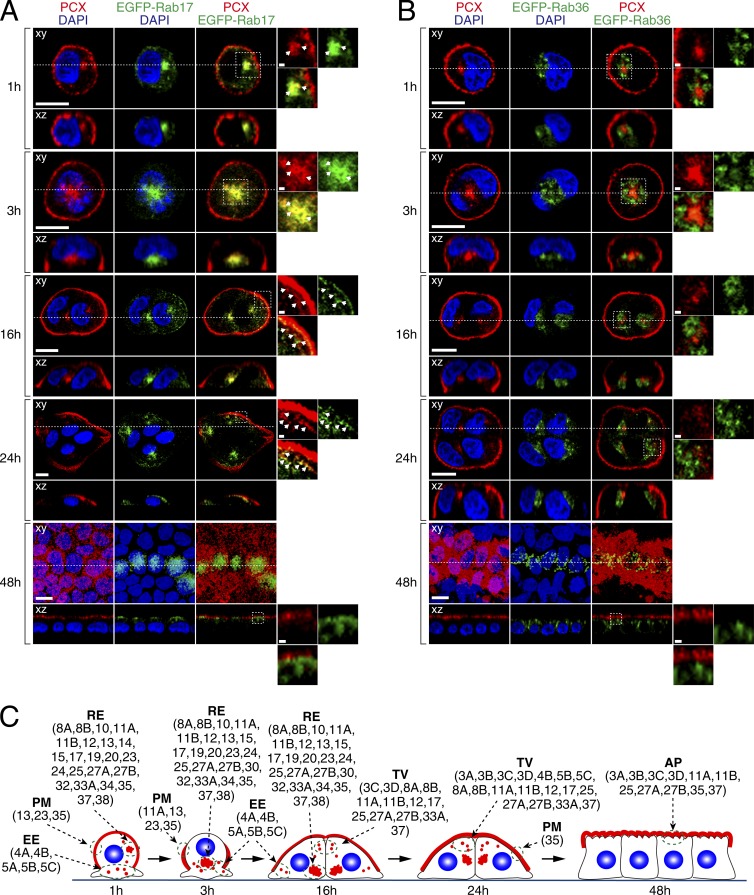
Colocalization of endogenous PCX with EGFP-tagged Rab17 and Rab36 in 2D MDCK II cells. MDCK II cells were transfected with plasmids carrying EGFP-tagged Rab17 (A) or Rab36 (B), plated on glass-bottom dishes, and fixed with PFA at the times indicated (see also Fig. S3). The cells were then stained with anti-PCX antibody (red) and DAPI (blue). The confocal xy section (top) and the xz section (bottom), which corresponds to the dashed line in the xy section, are shown. The fourth columns show magnifications of the boxed regions in the third columns. Bars: 10 µm; (insets) 1 µm. Note that EGFP-Rab17 and PCX were well colocalized at recycling endosomes just near the nucleus 1–3 h after plating and that their colocalization was also observed just beneath the plasma membrane (indicated by arrows) 24 h after plating. In contrast, no colocalization between EGFP-Rab36 and PCX was observed during PCX trafficking. (C) Schematic summary of the Rabs that colocalize with PCX (red) at specific membrane compartments (green doted circles) in 2D MDCK II cells (see also Table 1 and Fig. S3 for details). AP, apical plasma membrane; EE, early endosome; PM, plasma membrane; TV, transport vesicle.

