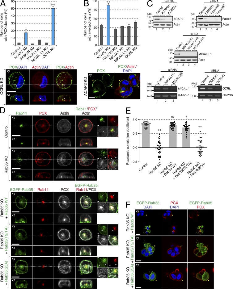
Figure 7.
Rab35 utilizes different effectors in 2D and 3D culture conditions to regulate PCX trafficking. (A and B) MDCK II cells that had been treated with control siRNA or siRNA against indicated Rab35 effectors were plated on glass-bottom dishes (A) or Matrigel-coated glass slides (B) and fixed with PFA 3 h (A) or 24 h (B) after plating. The cells were then stained with anti-PCX antibody (green), Texas red–conjugated phalloidin (red), and DAPI (blue). Values represent the mean and SEM of at least three independent experiments (n > 100 in each experiment). Significance was determined by one-way analysis of variance with Dunnett's post-test at 95% confidence interval. *, P < 0.05; **, P < 0.01. Bottom images show typical OCRL KD cells (A) and ACAP2 KD cells (B). (C) KD efficiency of siRNAs against Rab35 effectors as revealed by immunoblotting with specific antibody against each effector. siRNAs against ACAP2, Fascin, MICAL1, MICAL-L1, and OCRL were transfected into MDCK II cells. For ACAP2, Fascin, and MICAL-L1 the level of endogenous protein expression was detected with specific antibodies. For MICAL1 and OCRL the level of gene expression was determined by RT-PCR. Underlined siRNAs were used for the KD experiments. (D) Rab35-KO MDCK II cells were transfected with plasmids carrying EGFP-tagged wild-type (WT) Rab35, a Rab35(TS/TA) mutant with decreased affinity for ACAP2, or a Rab35(S5A) mutant completely unable to bind ACAP2 and with decreased affinity for other Rab35 effectors (Etoh and Fukuda, 2015). Control cells, Rab35-KO cells, and transfected Rab35-KO cells were then plated on glass-bottom dishes and fixed with PFA 3 h after plating. Control and Rab35-KO cells were stained with anti-Rab11 antibody (green), anti-PCX antibody (red) and Alexa Fluor 633 phalloidin (white). The transfected Rab35-KO cells were stained with anti-Rab11 antibody (red) and anti-PCX antibody (white). (E) The graph shows Pearson’s correlation coefficient of colocalization between PCX and Rab11 for cells shown in D. PCCs were calculated for at least 30 cells from three independent experiments. Significance was determined by one-way analysis of variance with Dunnett's post-test at 95% confidence interval. *, P < 0.05; **, P < 0.01; ns, nonsignificant. (F) Rab35-KO MDCK II cells were transfected with plasmids carrying EGFP-tagged Rab35 WT, the Rab35(TS/TA) mutant, or the Rab35(S5A) mutant. The cells were then plated on Matrigel-covered glass slides, fixed with PFA 24 h after plating, and stained with anti-PCX antibody (red) and DAPI (blue). Note that in 2D cells endosomal localization of PCX can be rescued by wild-type Rab35 and to some extent Rab35(TS/TA) mutant, whereas in 3D cysts, the inverted phenotype can only be rescued by wild-type Rab35. The white arrows, white arrowheads, and yellow arrowheads (D and F) point to RE-localized PCX, non-RE intracellular PCX, and peripheral PCX, respectively. For cells growing on Matrigel (B and F), the confocal xy section is shown. For cells growing on glass-bottom dishes (A and D), the confocal xy section (top) and the xz section (bottom), which corresponds to the dashed line in the xy section, are shown. Bars: 10 µm; (insets) 1 µm.

