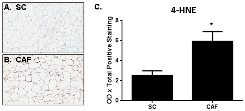
Figure 2. Oxidative stress is increased in WAT from CAF diet-exposed rats.
Anti-4-HNE IHC was performed in WAT collected from rats fed (A) SC or (B) CAF diet for 15 weeks. Representative 20× images are shown. C. Quantification of mean optical density x total positive staining ± SEM. N = 3. *p-value = 0.03.

