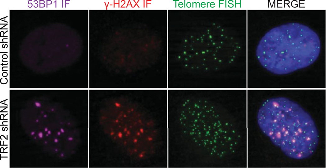
Figure 12.40.1.
In this example, HT1080 6TG cells that were untreated or transduced with a TRF2 shRNA are stained with a telomere PNA (green), γ-H2A.X (red), and 53BP1 IF (magenta), and DAPI (blue). Telomere dysfunction induced foci (i.e., TIF) are evident as co-localized IF and FISH signals in the TRF2 shRNA treated cell.

