
Figure 1.
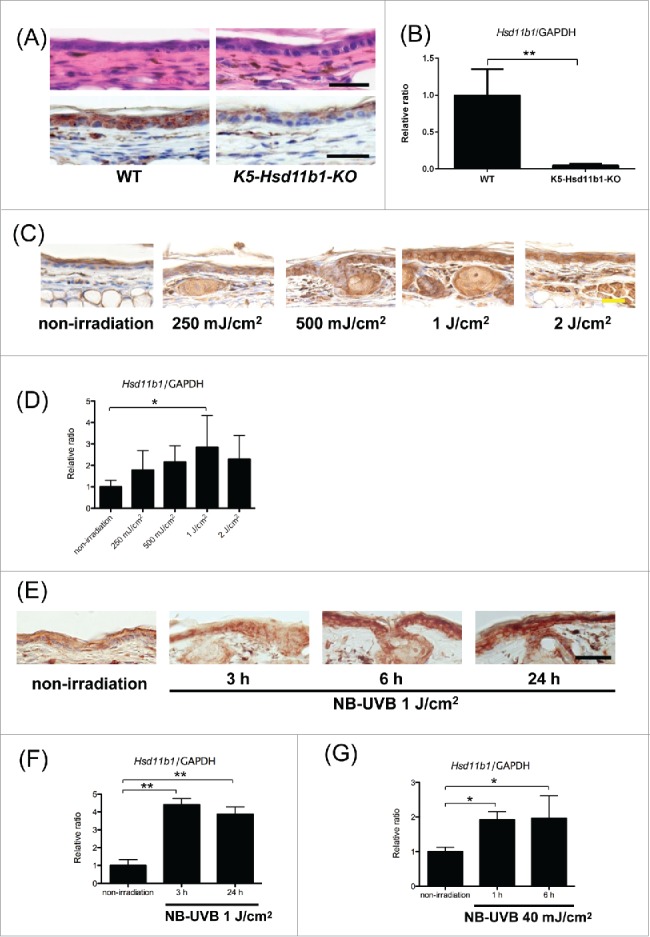
(For figure legend, see page 6.) Figure 1 (see previous page). Generation of K5-Hsd11b1-KO mice. The expression of 11β-HSD1 in the skin was enhanced following NB-UVB irradiation in WT mice. (A) Representative H&E staining (upper panel) and 11β-HSD1 staining (lower panel) of 2-month-old WT and K5-Hsd11b1-KO mouse skin Bar = 50 μm. (B) The relative expression levels of 11β-HSD1 in keratinocytes derived from WT and K5-Hsd11b1-KO mice were assessed using RT-PCR. GAPDH was used as an internal control. The bars indicate the mean ± SD (N = 5; *P < 0.05, Student's t-test). (C) Expression of 11β-HSD1 in 2-month-old WT mice irradiated with 250 mJ/cm2, 500 mJ/cm2, 1 J/cm2, and 2 J/cm2 NB-UVB. Skin specimens were harvested 24 hours after NB-UVB irradiation. Rabbit IgG was used as isotype control. Bar = 20 μm (N = 6 per group). (D) The relative expression levels of 11β-HSD1 in WT mice irradiated with 250 mJ/cm2, 500 mJ/cm2, 1 J/cm2, and 2 J/cm2 NB-UVB were assessed using RT-PCR. Skin samples were collected 24 hours after NB-UVB irradiation. GAPDH was used as an internal control. The bars indicate the mean ± SD (N = 6; **P<0.01, one-way ANOVA followed by the Bonferroni-Dunn test for multiple comparisons). (E) Expression of 11β-HSD1 in 2-month-old WT mice irradiated with 1 J/cm2 of NB-UVB. Skin specimens were harvested 3, 6, and 24 hours after NB-UVB irradiation. Rabbit IgG was used as isotype control. Bar = 50 μm (N = 6 per group). (F) The relative expression levels of 11β-HSD1 in WT mice irradiated with 1 J/cm2 NB-UVB were assessed using RT-PCR. Skin samples were collected at the indicated time points for unirradiated mice and 3 and 24 hours after NB-UVB irradiation. GAPDH was used as an internal control. The bars indicate the mean ± SD (N = 6; **P < 0.01, one-way ANOVA followed by the Bonferroni-Dunn test for multiple comparisons). (G) The relative expression levels of 11β-HSD1 in WT mouse-derived keratinocytes after NB-UVB irradiation with 40 mJ/cm2 NB-UVB were assessed using RT-PCR. Keratinocytes were collected at the indicated time points for unirradiated mice and 1 and 6 hours after NB-UVB irradiation. GAPDH was used as an internal control. The bars indicate the mean ± SD (N = 6; *P < 0.05, one-way ANOVA followed by the Bonferroni-Dunn test for multiple comparisons).