
Figure 3.
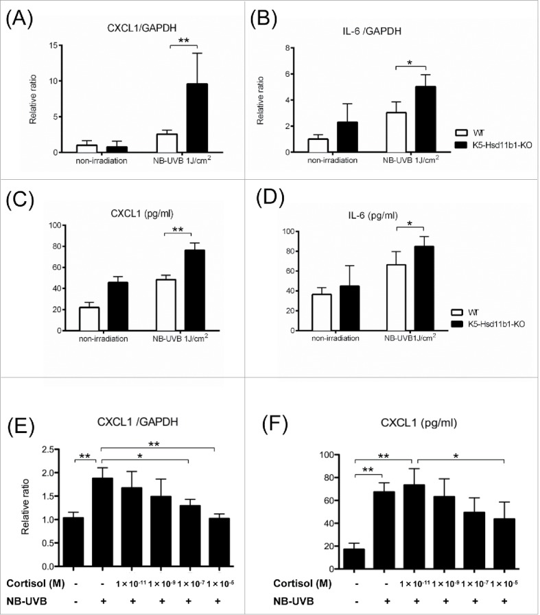
Inflammatory response to NB-UVB irradiation was enhanced in the K5-Hsd11b1-KO mouse skin. (A, B) Relative expression levels of CXCL1 (A) and IL-6 (B) in skin from 2-month-old mice 3 hours after NB-UVB irradiation at 1 J/cm2. GAPDH was used as an internal control. The bars indicate the mean ± SD (N = 6; **P < 0.01, *P < 0.05, two-way ANOVA followed by the Bonferroni-Dunn test for multiple comparisons). (C, D) The levels of CXCL1 (C) and IL-6 (D) in the skin of 2-month-old WT mice 10 hours after NB-UVB irradiation at 1 J/cm2 were measured using ELISA. The bars indicate the mean ± SD (N = 6; **P < 0.01, *P < 0.05, two-way ANOVA followed by the Bonferroni-Dunn test for multiple comparisons). (E) The indicated dose of cortisol was added to the cultured media of the WT mice-derived keratinocytes for 24 hours, and then cells were treated with or without 40 mJ/cm2 NB-UVB irradiation. Cells were harvested after 1 hour, and CXCL1 expression was investigated by rtPCR. GAPDH was used as an internal control. The bars indicate the mean ± SD (N = 6; **P < 0.01,*P < 0.05, one-way ANOVA followed by the Bonferroni-Dunn test for multiple comparisons). (F) The indicated dose of cortisol was added to the cultured media of WT mice-derived keratinocytes for 24 hours, and then cells were treated with or without 40 mJ/cm2 NB-UVB irradiation. Culture media was collected 10 h later, and concentrations of CXCL1 were measured by ELISA. The bars indicate the mean ± SD (N = 6; **P < 0.01, *P < 0.05, one-way ANOVA followed by the Bonferroni-Dunn test for multiple comparisons).