
Figure 4.
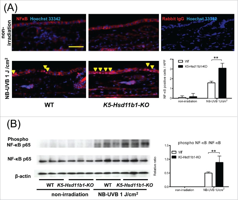
NB-UVB-induced activation of NF-κB in the K5-Hsd11b1-KO mouse skin was enhanced. (A) Immunofluorescence staining of NF-κB p65 (red) and nuclei (Hoechst 33342, blue) in skin of 2-month-old WT and K5-Hsd11b1-KO mice 3 hours after NB-UVB irradiation at 1 J/cm2. Rabbit IgG was used as isotype control. Bar = 100 μm. Three sections from each mouse were evaluated. The bars indicate the number of NF-κB p-65-positive cells translocated into the nucleus after NB-UVB irradiation (mean ± SD; N = 6; **P < 0.01, two-way ANOVA followed by the Bonferroni-Dunn test for multiple comparisons). (B) Western blot analysis of the phospho-NF-κB p65 expression in skin from 2-month-old WT and K5-Hsd11b1-KO mice 1 hour after NB-UVB irradiation at 1 J/cm2. The intensity of the bands was quantified using the NIH Image J software program. The phospho-NF-κB p65/NF-κB p65 was calculated. The bars indicate the mean ± SD (N = 3; *P < 0.05, 2-way ANOVA followed by the Bonferroni-Dunn test for multiple comparisons).