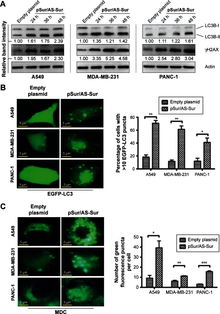
Figure 4.
Delivery of pSur/AS-Sur modulates autophagy in cancer cells.
Notes: (A) Cancer cells were transfected with either empty plasmid or pSur/AS-Sur for the indicated durations. The conversion of LC3B-II and the expression of γH2AX were examined by Western blotting. Equal protein loading was verified by actin. The numbers under each blot are the intensities of the blot relative to that of the control (empty plasmid). (B) Cancer cells were transfected with EGFP-tagged LC3B with or without pSur/AS-Sur cotransfection for 48 hours. LC3 puncta formation in cells (~200 cells) was observed under a fluorescence microscope. The number of puncta present in cells was analyzed using the ImageJ software. (C) Cancer cells were transfected with pSur/AS-Sur for 48 hours and then stained with MDC. The number of puncta present in cells (~200 cells) was analyzed using the ImageJ software. “*,” “**,” and “***” denote statistical significance with P-values <0.05, <0.01, and <0.001, respectively, between the testing groups.
Abbreviations: h, hours; MDC, monodansylcadaverine; pSur/AS-Sur, survivin promoter-driven antisense survivin.

