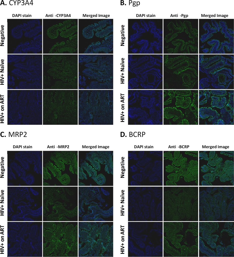
FIG 3.
Immunohistochemistry analysis of protein expression of selected intestinal metabolic enzymes and drug efflux transporters. To detect protein expression and localization, paraffin-embedded jejunal tissue biopsy specimens from each subject, fixed in 4% paraformaldehyde and mounted onto glass slides, were immunostained with anti-CYP3A4 mouse polyclonal antibody from Sigma-Aldrich (Oakville, Ontario, Canada) at a 1:600 dilution (A), anti-Pgp mouse monoclonal D-11 antibody from Santa Cruz Biotechnology, Inc. (Dallas, TX) at a 1:100 dilution (B), anti-MRP2 mouse monoclonal M2III-6 antibody from Kamiya Biomedical Company (Seattle, WA) at a 1:250 dilution (C), or anti-BCRP rat monoclonal BXP-21 antibody from Abcam Inc. (Toronto, Ontario, Canada) at a 1:100 dilution (D). After washing, each tissue slice was immunostained with the corresponding Alexa-488-conjugated secondary antibodies, i.e., donkey anti-rat IgG for BCRP or donkey anti-mouse IgG for all other proteins (Life Technologies Inc., Burlington, Ontario, Canada). Standard DAPI staining was used to identify cell nuclei. For each gene of interest, the entire set of tissue slices was immunostained in a single experiment with all steps performed simultaneously, and images were obtained at constant exposure, zoom, and background settings using a Zeiss LSM700 confocal microscope.

