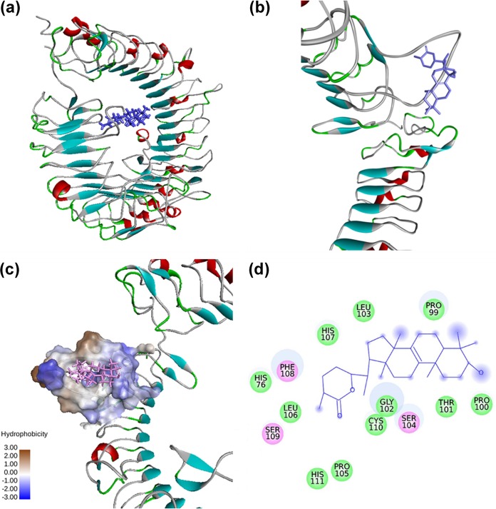
FIG 6.
Interaction between mouse TLR9 and astrakurkurone, as predicted by molecular docking. (a) Mouse TLR9-astrakurkurone docked complex. (b) Closer view of docked complex. (c) The surface of the mouse TLR9 binding site. (d) Intermolecular interaction of TLR9 with astrakurkurone. Residues involved in electrostatic and Van der Waals interactions are represented by pink and green circles, respectively. The solvent-accessible surface of an interacting residue is represented by a blue halo around the residue.

