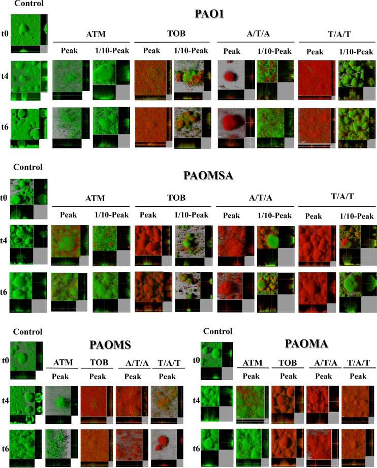
FIG 6.
Three-dimensional images and transverse sections of GFP (green)-tagged laboratory strain biofilms treated with peak (700 mg/liter ATM and 1,000 mg/liter ATM) and 1/10-peak concentrations (70 mg/liter ATM and 100 mg/liter TOB) and stained with propidium iodide (red). The images obtained at three time points (t0, t4, and t6) are shown.

