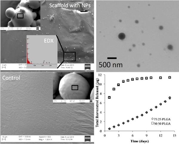
Figure 3.
Scaffold characterization and resveratrol release profile. (A) SEM image of aresveratrol nanoparticle-incorporated scaffold showing the nanoparticles on the surface of the sintered microsphere scaffolds PLGA. (B) SEM image of a blank PLGA scaffold (control) demonstrating a smooth surface without nanoparticles. (C) TEM image of individual resveratrol encapsulated nanoparticles. The approximate diameter of nanoparticles was 250 nm. (D) Resveratrol release profile from the scaffolds demonstrated a slower and more linear release when encapsulated with a higher molecular weight of PLGA.

