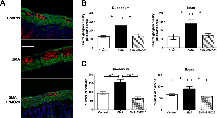
Fig 4. Enteric neurons in SMA mouse small intestine.
(A) Representative image of enteric neurons/ganglions in Duodenum myenteric plexuses from control, SMA and PMO25 treated SMA mice. Enteric neurons were stained with neuronal marker PGP9.5 (red). The muscular layer was stained with α-smooth muscle actin (green). Cell nuclei were stained with DAPI (blue). (B) Relative ganglion density in 3 groups of mice. Pixels of PGP9.5 immunostaining per captured field was used to quantify the ganglion density using imageJ software and expressed as pixels per unit area. Ganglion density was significantly increased in SMA mice in both duodenum (P = 0.028 vs control, N = 4 per group) and ileum (P = 0.018 vs control, N = 4 per group) and significantly decreased after PMO25 treatment (P = 0.038 in duodenum; P = 0.019 in ileum; N = 4 per group). (C) The mean number of neurons was also significantly increased in both duodenum (P = 0.0045 vs control, P< 0.001 vs PMO25 treatment) and ileum (P = 0.04 vs control, P = 0.012 vs PMO25 treated SMA) in SMA mice and was reduced significantly by PMO25 treatment (N = 6–8, * P < 0.05; ** P < 0.01; *** P < 0.001). Scale bar = 25 μm.

