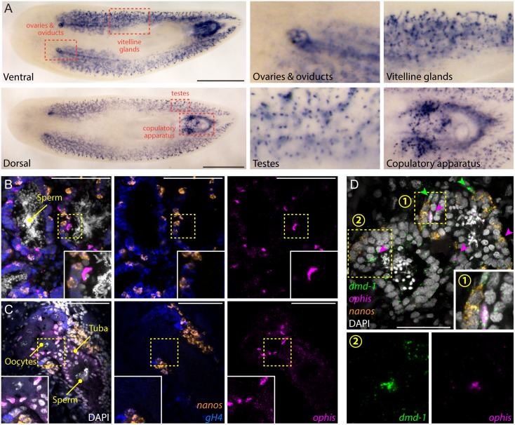
Fig 6. ophis is expressed in the somatic gonadal niche.
(A) Colorimetric ISH shows expression of ophis in somatic reproductive structures. Insets show magnified view of specific tissues indicated by red dashed boxes. (B and C) Triple-FISH labeling ophis (magenta), gH4 (blue), and nanos (orange). Within gonads, ophis expression is exclusive to somatic cells in the periphery of testis lobes (B) and in presumptive follicular cells of the ovaries (C). (D) Triple-FISH labeling ophis (magenta), nanos (orange), and dmd-1 (male somatic gonad cells, green) in the testes. ophis and dmd-1 are co-expressed inside testes (magenta arrowheads). dmd-1+/ophis- cells can be seen outside the testes (green arrowheads). Insets 1 and 2 show magnification of regions indicated by numbered yellow dashed boxes. DAPI (grey) labels nuclei in B–D. Scale bars are 1 mm in A, 100 μm in B and C, and 50 μm in D.

