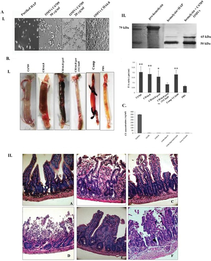
FIG 4.
Protease-associated OMVs are biologically active. (A) Biological activities of HAP-associated OMVs. Panel I, Int407 cells were treated with OMVs from C6709 at 50 μg/ml and 20 μg/ml and OMVs from CHA6.8 for 24 h at 37°C. The higher concentration of C6709 OMVs showed a cell-rounding effect, whereas the lower concentration showed a cell-distending effect. No morphological changes were observed in CHA6.8. Panel II, immunoblot analyses against hemolysin antiserum with 10 μg purified hemolysin treated with HAP and C6709 OMVs and purified hemolysin as positive control. (B) Panel I, effect of OMVs of Vibrio cholerae strains in the mouse ileal loop assay. Twenty micrograms (100 μg/ml) of OMVs from wild-type strain C6709 and its knockout derivatives was introduced into ligated ileal loops of adult mice. The mice were sacrificed after 6 h, loops were excised, and fluid accumulation was calculated as the loop weight/length ratio. Images of mouse ileal loops treated with V. cholerae OMVs are shown. The graph shows a summary of the data, represented as mean ± SD (n = 5 mice per group). Variables were compared for significance using two-way analysis of variance and the Bonferroni test (*, P value between 0.01 and 0.05; **, P value between 0.01 and 0.001). Panel II, histopathological studies of mouse ileal loops. Twenty micrograms of OMV-treated ileal tissues was processed for histopathological analysis, and photomicrographs were taken at a magnification of ×40. A, PBS-treated ileal tissues showed normal villi with mucosal structure; B, a magnified image of ileal tissues treated with C6709 OMVs shows dilated and ruptured villi with accumulation of inflammatory cells in mucosal layers; C, CHA6.8 shows an altered villous structure with mild hemorrhage in the submucosa; D, CHA 6.8 ΔprtV OMV-treated ileal tissues show grossly disrupted villi with hemorrhage in all layers of the mucosa; E, ileal tissues treated with OMVs of strain CHA6.8 ΔprtV ΔVC1649 show an almost-normal villous structure with minimum hemorrhage in the mucosa, submucosa, and lamina propria; F, OMVs from the VC1649 complemented strain show accumulation of inflammatory cells and hemorrhage in mucosal layers. (C) CT-beads ELISA results for OMVs from the C6709, CHA6.8, CHA6.8 ΔprtV, CHA6.8ΔprtV ΔVC1649, and Comp VC1649 strains. Culture supernatant of strain N16961 was used as a positive control.

