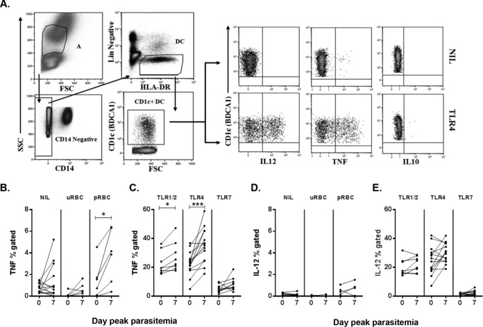
FIG 5.
CD1c+ mDC cytokine responsiveness to TLR or pRBC stimulation. (A) Representative staining of blood CD1c+ mDCs for intracellular cytokines. CD1c+ mDCs were identified as negative for lineage markers (CD14, CD3, CD19, and CD56), HLA-DR+, and CD1c+. Intracellular cytokine production by CD1c+ mDCs on day 0 (IL-12, TNF, and IL-10) under two conditions, ex vivo (NIL; top panel) and TLR4 (bottom panel), was determined. (B) TNF production on day 0 and day 7 ex vivo (NIL) and after uRBC or pRBC (P = 0.03) stimulation. (C) TNF production on day 0 and day 7 after TLR1/2 (P = 0.02), TLR4 (P = 0.0002), or TLR7 stimulation. (D) IL-12 production on day 0 and day 7 ex vivo (NIL) and after uRBC or pRBC stimulation. (E) IL-12 production on day 0 and day 7 after TLR stimulation. A Wilcoxon matched-paired test was used for comparison between days 0 and day 7. *, P < 0.05; ***, P = 0.0002. Line graphs show data for all subjects (n = 14; the exceptions include 8 individuals for TLR1/2, 10 individuals for TLR7, and 6 individuals for uRBCs and pRBCs). FSC, forward scatter; SSC, side scatter; uRBC, uninfected red blood cells; pRBC, parasitized red blood cells.

