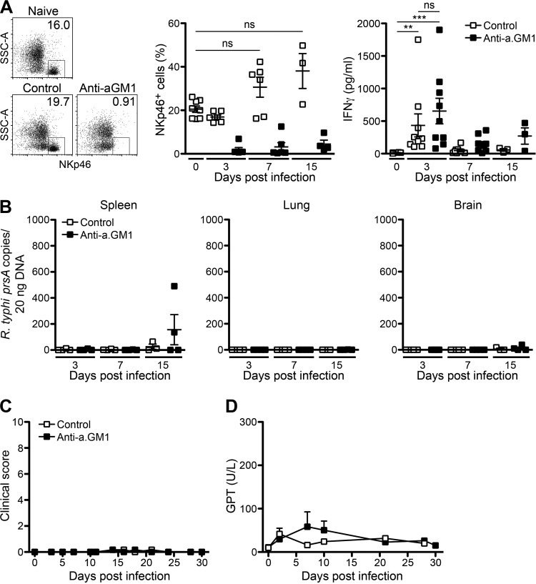
FIG 2.
Depletion of NK cells does not affect the course of disease in C57BL/6 RAG1−/− mice upon R. typhi infection. (A) C57BL/6 RAG1−/− mice were treated with anti-asialo GM1 antibody (Anti-a.GM1) or control serum every 5 days starting 1 day prior to infection with 2 × 10⁶ SFU R. typhi s.c. The percentages of NKp46+ NK cells in blood and plasma levels of IFN-γ were assessed by LEGENDplex assay. Representative results for the staining of blood cells from a naive mouse and R. typhi-infected mice that received either control serum or anti-asialo GM1 antibody are shown in the scatter plots (day 3 postinfection). Graphs show statistical analysis of the percentages of NKp46+ cells (y axis) and IFN-γ plasma levels (y axis) at indicated points in time postinfection (x axis). Each symbol represents the result for a single mouse; bars and whiskers show the mean and SEM. Statistical analysis was performed with the Kruskal-Wallis test. Asterisks indicate statistically significant differences (**, P < 0.01; ***, P < 0.001). SSC, side scatter. (B) Bacterial loads were determined by qPCR of prsA in spleen, lung, and brain tissues. R. typhi prsA copy numbers (y axis) at indicated points in time (x axis) are depicted. Each symbol represents the result for a single mouse. (C and D) The course of disease was evaluated by clinical scoring (n = 5 for each group) (y axis) (C) and quantification of GPT in the serum (n = 3 for each group) (y axis) (D) at the indicated points in time (x axes). Error bars show SEM.

