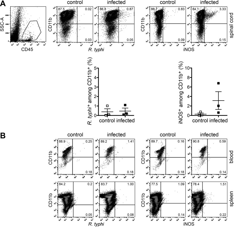
FIG 7.
Inflammatory macrophages are not detectable in the periphery. (A) Cells from the spinal cords of the same control and R. typhi-infected C57BL/6 RAG1−/− mice described in the legend to Fig. 6, taken at the time of death, were gated on CD45+ cells and further stained for CD11b, R. typhi, and iNOS as indicated (scatter plots). The graphs show the statistical analysis of these measurements. Each symbol represents the result for a single mouse; bars and whiskers show the mean and SEM. (B) In a similar manner, blood and spleen cells (as indicated on the right) from the same mice were stained for CD11b, R. typhi (left), and iNOS (iNOS). Representative results of staining for one mouse are depicted.

