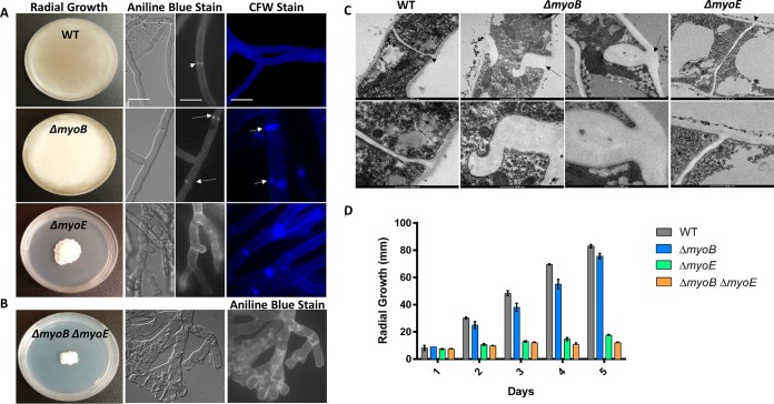
FIG 1.
Myosins are required for proper hyphal and septal morphology. (A) Deletion of myoB resulted in white colonies with wild-type (WT) radial growth. Deletion of myoE resulted in white, compact colonies. Conidia (104) were spotted onto GMM agar and incubated for 5 days at 37°C. Single deletion of myoB resulted in β-(1,3)-glucan mislocalization, and deletion of myoB or myoE resulted in chitin mislocalization. Conidia (104) were inoculated into 5 ml GMM on sterile coverslips for 18 h at 37°C. The coverslips were stained with aniline blue to stain β-glucan or calcofluor white (CFW) to stain chitin and were visualized using fluorescence microscopy. The arrowhead indicates a normal septum, the dashed arrows indicate β-(1,3)-glucan mislocalization, and the solid arrows indicate chitin mislocalization. Bar, 10 μm. (B) Strains with deletions of both myoB and myoE result in hyperbranching and β-(1,3)-glucan patches throughout the hyphae. (C) MyoB is required for proper septum formation. Deletion of myoB resulted in malformed septa thicker than in the wild type. Septa were visualized using TEM. The arrowheads point to normal septa, and the dashed arrows indicate aberrant septa in the myoB deletion strain. (D) Strains harboring a deletion of myoE resulted in a significant radial-growth defect. Conidia (104) were spotted onto GMM agar and incubated for 5 days at 37°C. Radial growth was measured every 24 h. The error bars indicate standard errors of the means.

