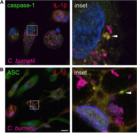
FIG 5.

Caspase-1 and ASC colocalize with IL-1β near avirulent C. burnetii. hAMs were infected with avirulent C. burnetii for 72 h and then processed for confocal fluorescence microscopy using antibodies directed against caspase-1 (A), ASC (B), and IL-1β and C. burnetii. DAPI was used to stain DNA (blue), and colors are shown in images. Bar, 10 μm. Caspase-1 and ASC colocalize with IL-1β (yellow; arrowheads) near C. burnetii, suggesting that inflammasomes are located near the PV for detection of avirulent bacteria.
