
FIG 2.
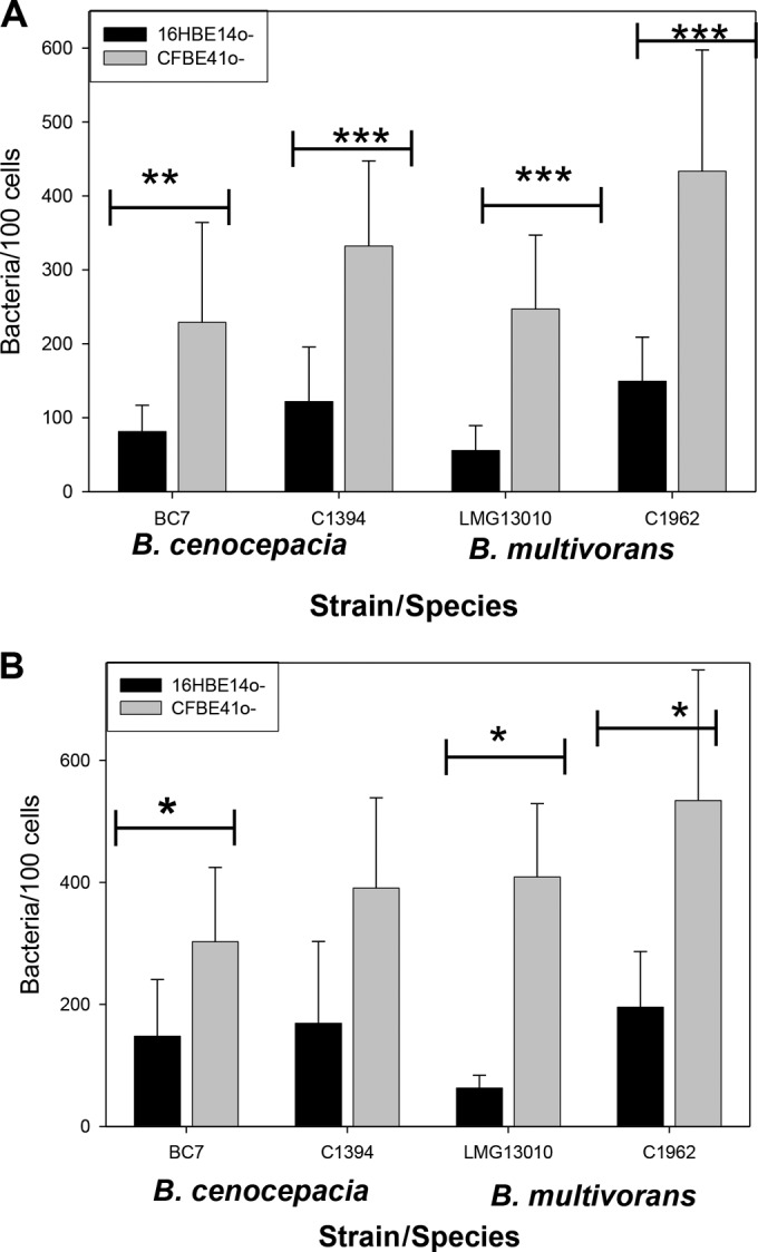
Attachment of Bcc to CFBE41o− and 16HBE14o− cells was examined by confocal microscopy at MOIs of 10:1 (A) and 50:1 (B). Attached bacteria were labeled with FITC-labeled antibody, and cells were stained with DAPI. Each bar represents means (± standard errors of the means) of data from two independent experiments, with 10 fields counted per strain. Statistical significance was determined by one-way ANOVA (***, P < 0.001; **, P < 0.005; *, P < 0.01).