FIG 4.
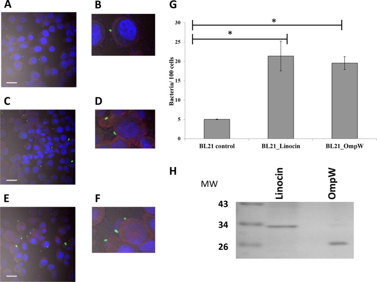
(A to F) Attachment of recombinant E. coli cells expressing either linocin (C and D) or OmpW (E and F) to CFBE41o− cells by confocal microscopy compared with E. coli BL21 controls (A). Bacteria (induced with 1 mM IPTG) were applied to CFBE41o− cells (MOI of 50:1) for 30 min and stained with FITC-conjugated anti-E. coli antibody, and epithelial cells were counterstained with phalloidin and DAPI and superimposed with differential interference contrast imaging. Bars = 20 μm. Panels B, D, and F show zoomed images to highlight bacteria interacting with epithelial cells. (G) Bacterial cell counts in 20 randomly selected fields for each strain, expressed as the number of bacteria attached per 100 epithelial cells, from two independent experiments. *, P < 0.05 as determined by one-way ANOVA. (H) Purified recombinant linocin and OmpW on a Coomassie blue-stained SDS-PAGE gel. MW, molecular weight (in thousands).

