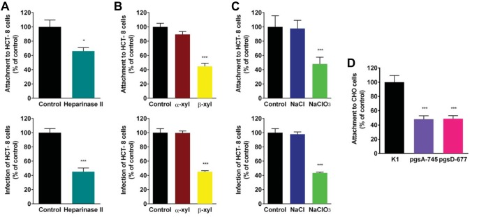
FIG 5.
Disruption of cell surface GAGs reduces C. parvum attachment and infection. (A) HCT-8 cells were treated with heparinase II or lyase buffer; this was followed by incubation with CFSE-labeled sporozoites or infection with oocysts. (B) HCT-8 cells were incubated with α-xyloside, β-xyloside, or medium alone and incubated with CFSE-labeled sporozoites or infected with oocysts. (C) HCT-8 cells were incubated with NaCl, NaClO3, or medium alone and incubated with CFSE-labeled sporozoites or infected with oocysts. (D) CHO K1, pgsA-745, and pgsD-677 cells were incubated with CFSE-labeled sporozoites. For all experiments, attachment and infection were quantified by fluorescence microscopy. Data represent the mean ± the SEM. *, P < 0.05; ***, P < 0.0005 (compared to the control [buffer or medium alone or CHO K1 cells]).

