Figure 1.
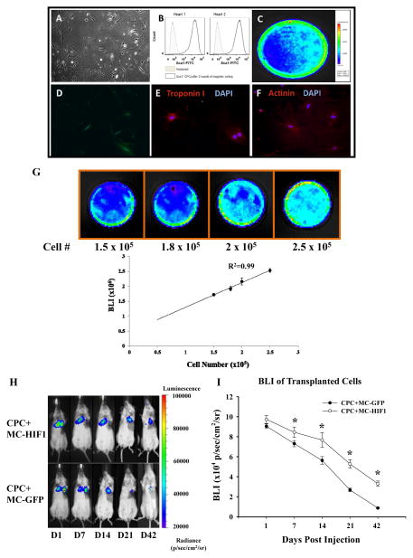
Co-delivery of hypoxia inducible-factor 1 (HIF-1) driven by minicircle (MC) plasmid promotes survival of transplanted Sca1+ cardiac progenitor cells (CPCs) in the ischemic heart. (A) The morphology of isolated CPCs growing on gelatin coated dish. (B) Flow cytometric analysis of purified Sca1+ CPC population from two preparations. Typical purity of isolation is >95%. CPCs isolated from transgenic mice express robust (C) firefly luciferase (Fluc) and (D) GFP expression. After culturing in cardiac differentiation induction medium, differentiated CPCs stained positively with (E) cardiac Troponin I and (F) α-actinin demonstrating its amenability to lineage commitment. (G) CPCs were seeded into dishes with increasing cell numbers. Assessment of BLI signals showed a robust linear correlation (R2=0.99) between the cell number and Fluc expression, which is crucial for accurate tracking of cell survival by in vivo imaging. (H) Representative BLI of animals injected with CPCs intramyocardially together with either MC-GFP or MC-HIF1 following myocardial infarction (MI) at indicated time points. (I) Quantitative analysis of longitudinal BLI signal demonstrates that CPCs co-delivered with MC-HIF1 had better survival compared to CPCs co-delivered with MC-GFP over a period of 42 days. *P<0.05 (N=10/group).

