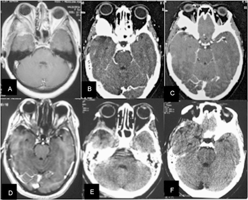
Fig. 1.

Sphenoorbital meningioma. (A–C) Preoperative magnetic resonance imaging (MRI) and computed tomography (CT) scan demonstrating a right sphenoorbital recurrence. (D–F) Postoperative MRI and CT scan demonstrating radical removal of the dura and bone involvement by the tumor.
