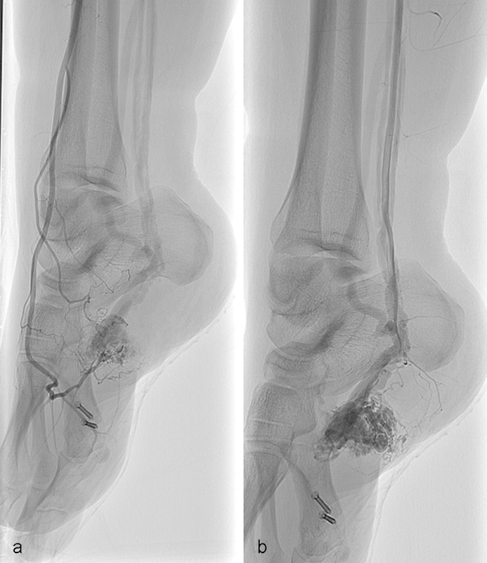
Fig. 3.

Diagnostic angiogram of the right foot. (a) Selective angiogram of the anterior tibial artery shows shunting and rapid egress of contrast from the arteriovenous malformation. (b) Selective angiogram of the posterior tibial artery similarly demonstrates rapid shunting and egress of contrast from the lesion.
