
Fig. 13.
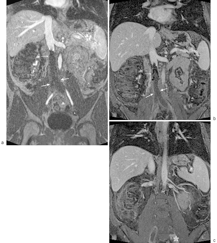
MR venography with occlusive IVC thrombosis extending from an IVC filter. (a) Venous phase contrast-enhanced MRA using gadofosveset trisodium demonstrates occlusive IVC and bilateral iliac thrombus (thick arrows) extending inferiorly from an IVC filter (thin arrow). Note that the distal iliac vein on the left is not opacified, which could be either thrombus or secondary to prolonged arteriovenous transit (asterisk). (b) Steady-state imaging using a 3D, T1-weighted, fat-saturated acquisition demonstrates the IVC filter (thin arrow) with peripheral enhancement around the occlusive IVC and bi-iliac thrombus (thick arrows). (c) Steady-state, 3D, T1-weighted, fat-saturated image demonstrates the inferior-most extent of the left iliac thrombus (asterisk). The thrombus extends peripherally to the confluence of the internal and external iliac veins.