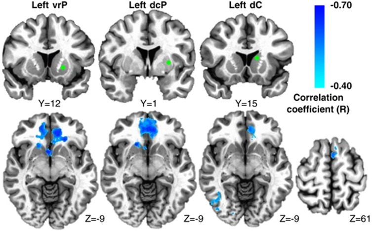
Figure 2.
Plasma C-reactive protein (CRP) was negatively associated with functional connectivity between ventral rostral putamen (vrP), dorsal caudal putamen (dcP) and dorsal caudate (dC) subdivisions of the striatum and other cortical brain regions in depressed subjects. Examples from the left hemisphere of functional connectivity between vrP, dcP and dC regions of interest (green seeds) and cortical brain regions that were negatively correlated with CRP (cyan-blue intensity; R=−0.40 to −0.70). Clusters in ventromedial prefrontal cortex (BA11; x=−6 to 2, y=27 to 36, z=−11 to −19; R=−0.62 to −0.53), right fusiform gyrus (BA37; x=37 and 35, y=−61 and −56, z=−14 and −16) and left superior frontal gyrus/supplementary motor area (BA6; x=−5, y=17, z=59; R=−0.51 to −0.53) are overlaid onto canonical structural brain images in the axial plane (z=−9 and 61), corrected P<0.05.

