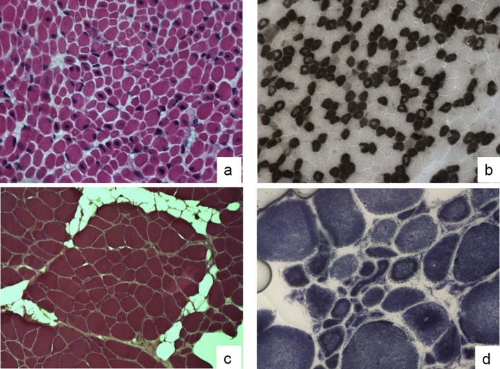
Fig. 1.
Muscle biopsies of cases IIIa and V. Muscle biopsy from patient IIIa: Hematoxylin–eosin stain (HE) (a) shows a muscle fiber size variability and many fibers (30–35%) contain a single central nucleus. ATPase 4.6 stain (b) reveals also a type 1 fiber predominance and atrophy; in particular, many fibers show a central area with a reduced myofibrillar reaction. A marked fiber size variation, several small fibers with internal nuclei and an increase of connective-adipose tissue have been observed in the HE stain (c) of a muscle biopsy from patient V. At NADH staining (d) several fibers display radial strands and internal dark ring necklace fibers.

