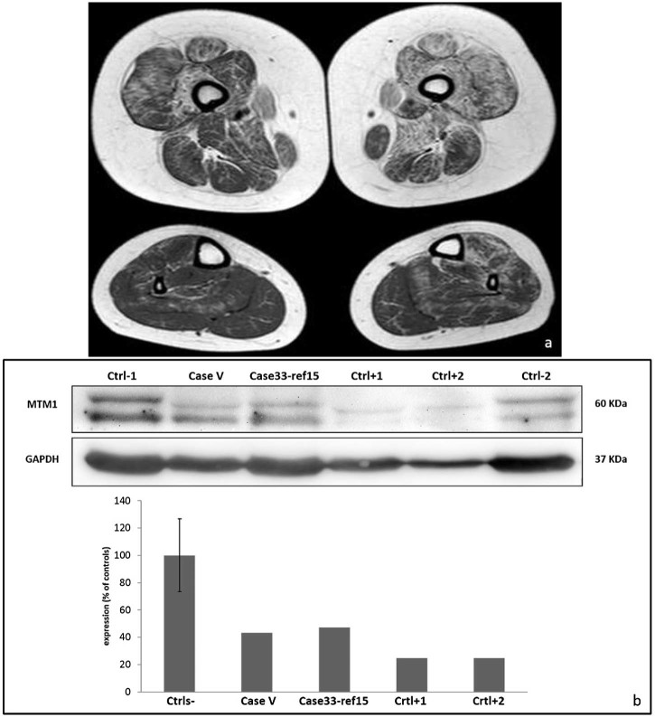
Fig. 2.
Imaging and WB of case V. In patient V (a), most muscles are affected asymmetrically (sn > dx) with only the adductor longus relatively spared. At leg level, the tibialis anterior and soleus muscles are affected on the left side. Immunolabeling and relative quantification of the MTM1 gene product (b) show a reduced band in patient V compared to the controls.

