
FIGURE 1.
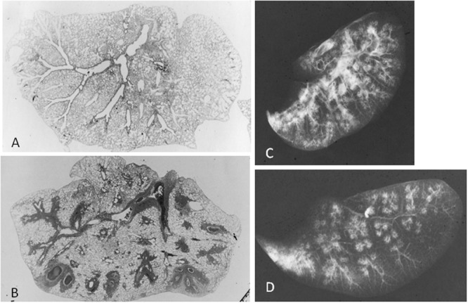
Mycoplasma pulmonis infected mice, 2 weeks after inoculation. Low-magnification photomicrographs of non-infected lung (A) and infected lung (B; HE x17). (C) Radiograph of inflated lung of infected mice reveals bronchovascular bundles thickening, nodules, and ground-glass attenuation. (D) Radiograph of thin-sliced lung of infected mice shows nodules with centrilobular distribution and consolidation. Reproduced with permission from Tanaka (2016).