
FIGURE 3.
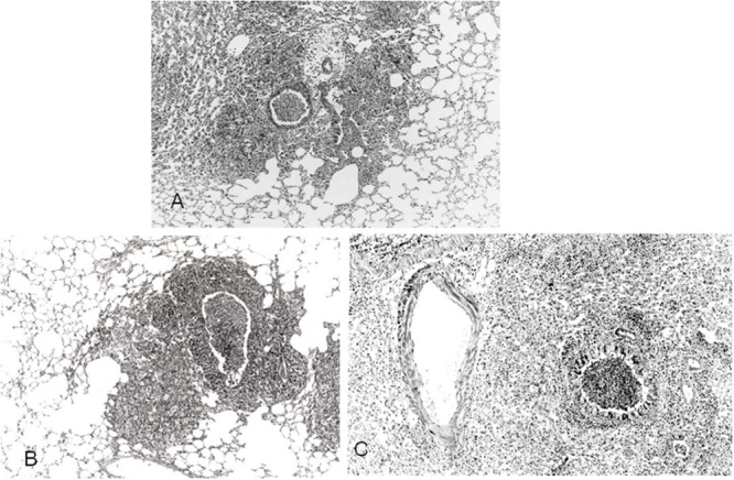
Pathological observation of lung from M. pulmonis infected mice (HE x170). (A) Mice without treatment. (B) Mice treated with interleukin-2, showing marked peribronchial lymphocyte cuffing and macrophage accumulation at the end bronchiole. (C) Mice treated with PSL, disclosing predominant intra-alveolar inflammatory cell Infiltration and faint perivascular lymphocyte cuffing. Reproduced with permission from Tanaka and Tamura (1989).