FIGURE 4.

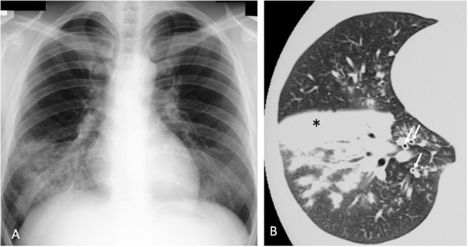
Mycoplasma pneumoniae pneumonia in human. (A) Chest x-ray shows infiltrates in the right lower lobe. (B) Consolidation (∗) and bronchovascular bundles thickening (↑) on CT scan. Reproduced with permission from Tanaka and Hayashi (2007).

Mycoplasma pneumoniae pneumonia in human. (A) Chest x-ray shows infiltrates in the right lower lobe. (B) Consolidation (∗) and bronchovascular bundles thickening (↑) on CT scan. Reproduced with permission from Tanaka and Hayashi (2007).