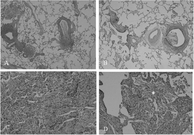
FIGURE 5.

Photomicrograph of open lung biopsy specimens in recovery phase of patients with M. pneumoniae pneumonia. Low-magnification views of small airways show cellular bronchiolitis and exudate in the lumen (A,B). High-magnification of alveolar area disclose stuffed alveoli with exudate, fibrin, neutrophil, and granulation tissue in alveolar duct (∗; C,D). Reproduced with permission from Tanaka (2016).
