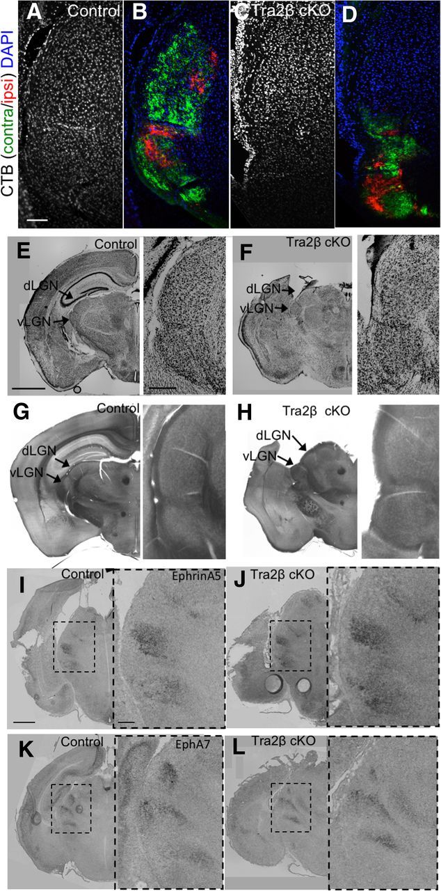
Figure 3.

Tra2β cKO LGN tissue is morphologically similar to that of control animals. A–D, 20 μm coronal sections of adult (P45–P80) animals stained with DAPI (blue) confirm the existence of cell nuclei in controls (A) and Tra2β cKO (C) visual thalamus, verified by labeling RGC axons originating from the left eye with CTβ-488 (green) and right eye with CTβ-555 (red) in control (B) and Tra2β cKO (D). E, F, NISSL-stained 50 μm sections of adult controls (E) and Tra2β cKO (F; arrows indicate locations of dLGN and vLGN) have similar staining patterns. G, H, Cytochrome oxidase staining of 50 μm sections of adult controls (G) and Tra2β cKO (H) arrows indicate the dLGN and vLGN have similar staining patterns. I–L, RNA in situ hybridization of coronal sections from P2 animals, dashed box indicates visual thalamus; Ephrin-A5 expression in control animals (I) and Tra2β cKO (J); EphA7 expression in controls (K) and Tra2β cKO (L) show similar patterns. Arrows indicate locations of dLGN and vLGN. Inset, Zoom of original images. Scale bars: A–D, 100 μm; E–H, 1 mm; inset, 200 μm; I–L, 500 μm; inset, 100 μm.
