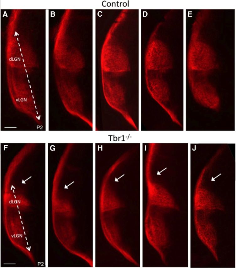
Figure 5.

Image forming RGCs fail to properly target the dLGN of Tbr1 KO animals. A–J, Coronal sections of the five P2 control mice (A–E) and five Tbr1 KO mice (F–J), that have had their contralateral RGC axons labeled with CTβ-555 (red). Distance of RGC innervation within LGN in control is significantly longer than mutant (dashed line), leaving the dorsal most dLGN lacking innervation (white arrows). Scale bar,100 μm.
