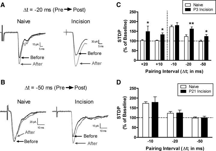
Figure 4.
Tissue damage during a critical period of early life widens the temporal window for t-LTP at primary afferent synapses onto adult spinal projection neurons. A, Examples of monosynaptic primary afferent-evoked EPSCs recorded in mature lamina I spino-parabrachial neurons from naive mice or mice that experienced hindpaw surgical incision at P3 both before (black) and after (gray) the administration of a Pre → Post pairing protocol at an interval of −20 ms. Note the greater magnitude of t-LTP in the incision group. Each displayed EPSC represents the average of 20 consecutive responses. B, Representative traces demonstrating that Pre → Post pairings at Δt = −50 ms failed to induce t-LTP in naive projection neurons, but could potentiate afferent-evoked EPSCs in mature projection neurons when preceded by neonatal incision. Illustrated EPSCs represent the average of 20 consecutive responses. C, Neonatal tissue damage broadened the timing window governing t-LTP at afferent synapses onto mature projection neurons because the potentiation of EPSC amplitude was significantly greater at Pre → Post pairings of −20 ms and −50 ms compared with naive littermate controls (n = 7–9 in each group; *p < 0.05; **p < 0.01; two-way ANOVA with Holm–Sidak multiple-comparisons test, right). Notably, whereas Post → Pre pairings at +10 ms and +20 ms intervals did not alter the efficacy of primary afferent synapses onto naive projection neurons (left), t-LTP was observed with these reverse pairings in adult projection neurons from neonatally incised mice (*p < 0.05 vs naive; two-way ANOVA with Holm–Sidak multiple-comparisons test). D, Delaying the surgical injury until P21 failed to evoke the same facilitation of t-LTP at Pre → Post pairings of −20 ms or −50 ms (n = 6–9 in each group; p = 0.81; two-way ANOVA), as was observed with P3 injury.

